فایل:Human brain anterior-inferior view description.JPG
Human_brain_anterior-inferior_view_description.JPG (۳۳۰ × ۴۷۵ پیکسل, فایل اؤلچوسو: ۳۱ کیلوبایت, MIME نوعو: image/jpeg)
قیساسی
| آچیقلاما |
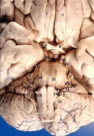
Human brain - anterior-inferior view - cerebral nerves I. N. olfactorius - The fila olfactoria (approximately 20 nerve rootlets on each side) comprise the Olfactory nerves (I) which run from the olfactory receptors in the nasal cavity to the olfactory bulb. II. N. opticus - The Optic nerves (II) are large & join at the midline to form the optic chiasm, then continue laterally as the optic tracts. The optic nerve is not a true nerve but rather a CNS tract. III. N. oculomotorius - The Oculomotor nerves (III) emerge from a depression in the midbrain, the interpeduncular fossa, just caudal to the optic chiasm. IV. N. trochlearis - The Trochlear nerves (IV) are small & are seen on the lateral surface of the midbrain. They are the only nerves which exit from the posterior side of the brain. V. N. trigeminus - The Trigeminal nerves (V) are large and emanate from the lateral surface of the pons. VI. N. abducens - The Abducens nerves (VI) exit near the midline from the inferior pontine sulcus which separates the pons from the medulla. VII. N. facialis - Moving laterally in the inferior pontine sulcus, the Facial nerves (VII) can be seen. VIII. N. vestibulocochlearis - Slightly lateral to the facial nerve is the Vestibulocochlear nerve (VIII). IX. N. glossopharyngeus - The postolivary sulcus is a groove running rostrocaudally on the lateral surface of the medulla. From this sulcus pass the small Glossopharyngeal nerves (IX) rostrally and... X. N. vagus - ...the much larger Vagus nerves (X) caudally. XI. N. accessorius - The Spinal Accessory nerves (XI) exit the cervical cord then pass rostrally through the foramen magnum to exit the cranial vault with the lossopharyngeal and vagus nerves. XII. N. hypoglossus - The Hypoglossal nerves (XII) exit the medulla via the preolivary sulcus.
|
| تاریخ | |
| قایناق | http://www.healcentral.org/healapp/showMetadata?metadataId=40566 (Internet Archive of file description page) |
| یازار |
John A Beal, PhD Dep't. of Cellular Biology & Anatomy, Louisiana State University Health Sciences Center Shreveport |
| ایجازه (فایلین تکرار ایستیفادهسی) |
CC-BY |
| آیری نوسخهلر |
 |
لیسانس
- سیزین ایجازه نیز وار دیر:
- بؤلۆشمک – اثرین کپی، پایلاشماق و چاتدیرماسی اوچون
- تلفیق ائت – اثرین توتوشدورماسی اوچون
- آشاغی داکی شرایطله:
- تخصیص – اثری مؤلف و یا لیسئنزییالی طرفیندن گؤستریلدیی کیمی ایستیناد ائتملیسینیز (آنجاق سنی دستکلدیینی و یا اثردن ایستیفاده ائتمیینیزی تکلیف ائتمملیسینیز).
این پرونده، که در اصل در http://www.healcentral.org/healapp/showMetadata?metadataId=40566 ارسال شده بود، در ۲۵ سپتامبر ۲۰۱۳ توسط بازبین Eleassar بررسی شد. این بازبین تأیید کرد که پرونده در آن محل و در آن تاریخ تحت مجوز اعلامشده در دسترس بودهاست.
|
فايل گئچمیشی
فايلین اولکی وئرسیياسینی گؤرمک اۆچون گۆن/تاریخ بؤلمهسیندهکی تاریخلری تێقلايین.
| تاریخ/واخت | کیچیک عکس | اؤلچولر | ایشلدن | باخیش | |
|---|---|---|---|---|---|
| ایندیکی | ۲۴ ژوئن ۲۰۰۶، ساعت ۱۸:۲۲ | | ۳۳۰ در ۴۷۵ (۳۱ کیلوبایت) | Patho | {{Information| |Description='''Human brain - anterior-inferior view - cerebral nerves''' I. N. olfactorius - The fila olfactoria (approximately 20 nerve rootlets on each side) comprise the Olfactory nerves (I) which run from the olfactory receptors in |
فايل ایشلدمهسی
آشاغیداکی صحیفه بو فایلا باغلانیر
فایلین باشاباش ایشلنیلمهسی
بو فایل آشاغی داکی آیری ویکی لرده ایشلَنیر:
- ar.wikipedia.org-ده ایشلنمهسی
- az.wikipedia.org-ده ایشلنمهسی
- bs.wikipedia.org-ده ایشلنمهسی
- de.wikipedia.org-ده ایشلنمهسی
- de.wikibooks.org-ده ایشلنمهسی
- en.wikipedia.org-ده ایشلنمهسی
- Cranial nerves
- User talk:Hovea
- User talk:Wouterstomp
- User talk:Nephron
- User talk:NCurse
- User talk:Robotsintrouble
- User talk:Was a bee
- User talk:Bloomingdedalus
- User talk:Read-write-services
- User talk:Attys
- User talk:Bakerstmd
- User talk:Manfi
- User talk:Mikepascoe
- User talk:Vokesk
- User talk:Neuraxıs
- User talk:Cmungall
- User talk:Qxukhgiels
- User talk:DocElisa
- User talk:JakobSteenberg/Archives/1
- User talk:Meteor sandwich yum
- User talk:Slashedone
- User talk:Anindya07
- User talk:Mattimussi
- User talk:Jelly Bean MD
- User talk:Benrusholme
- User talk:Brad.w.english
- User talk:Krsna ss
- User talk:Tom (LT)/Archive 4
- User talk:Dregon131
- User talk:Sapientia42
- User talk:Tilifa Ocaufa
- User talk:Snow Rise/Archive 6
- User talk:Arcadian
- User talk:Outofbattery
- User talk:Anatomyczar
- Wikipedia:WikiProject Anatomy/Newsletter/4
- User talk:Supravibhatsupravi
- User talk:Athikhun.suw
- User talk:CFCF/Archive 6
- User talk:Lewisskinner/ArchiveJul 2015
- User talk:Tyrol5/Archive 6
- User talk:CFCF/Archive 7
- User talk:DiverDave/Archive 5
- User talk:Caledones/archive/archive4
بو فایلینسراسری ایستفاده لرینه باخین.